
Ghi công
Nguồn ảnh
Ảnh chụp thật từ Wikimedia Commons, kho lưu trữ của chính phủ Hoa Kỳ và các nguồn thuộc phạm vi công cộng hoặc được cấp phép mở khác.
| Ảnh | Tác giả | Giấy phép | |
|---|---|---|---|
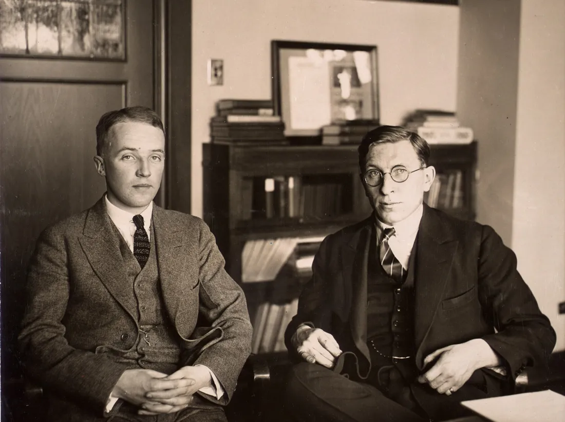 | C. H. Best and F. G. Banting ca. 1924 Insulin P10103 0001 | Unknown author | Public domain ↗ |
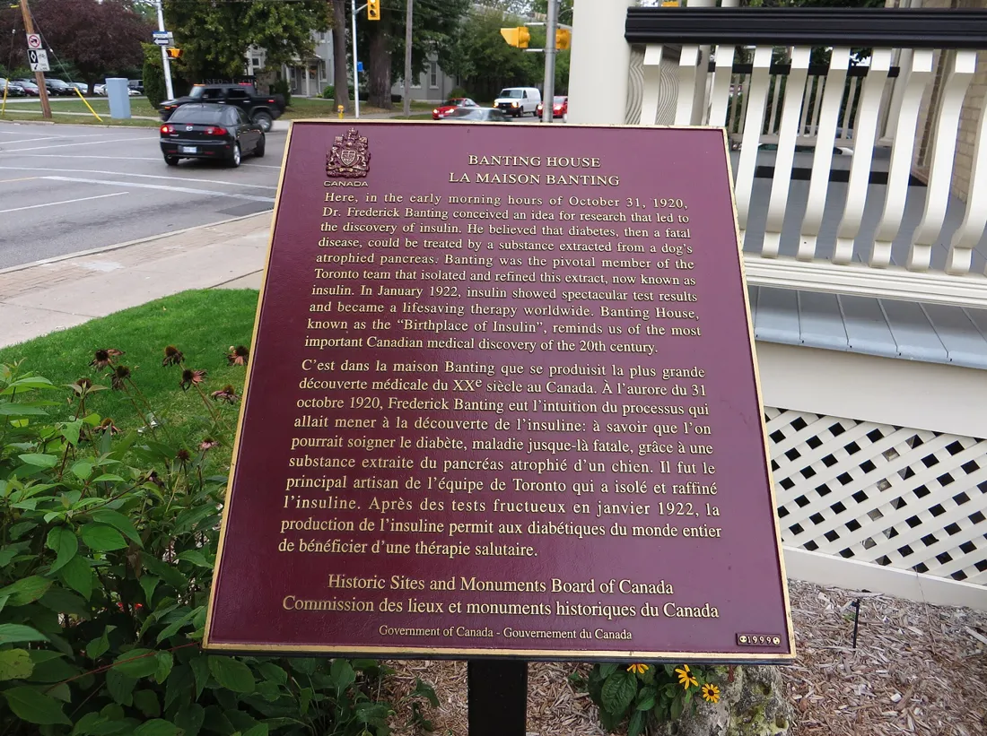 | Banting House, Birthplace of Insulin, London, Ontario (21650793378) | Ken Lund from Reno, Nevada, USA | CC BY-SA 2.0 ↗ |
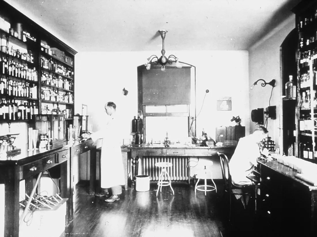 | Kinyoun's laboratory | Unknown photographer | Public domain ↗ |
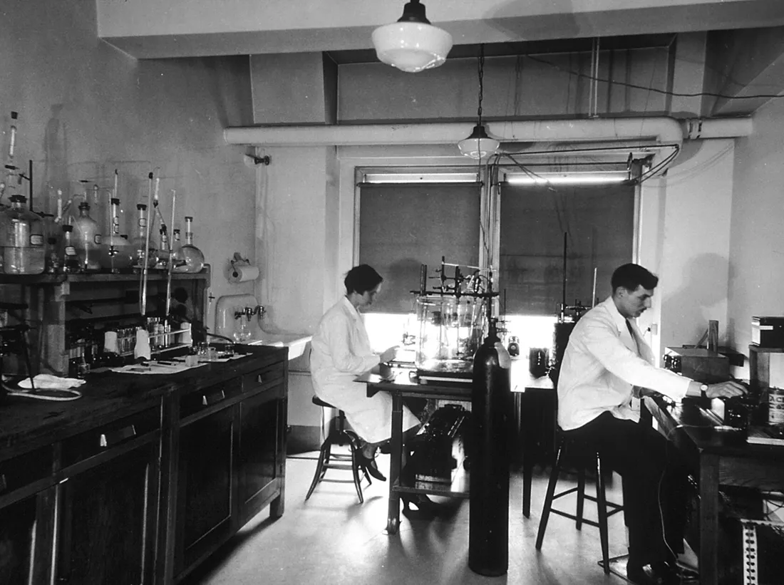 | Chemical laboratory at the central cancer research labs | Unknown photographer | Public domain ↗ |
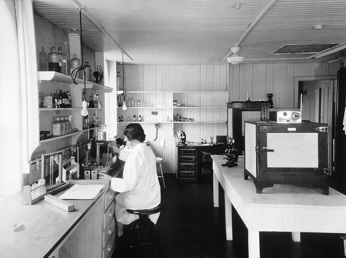 | Immunology laboratory at the central cancer research labs | Unknown photographer | Public domain ↗ |
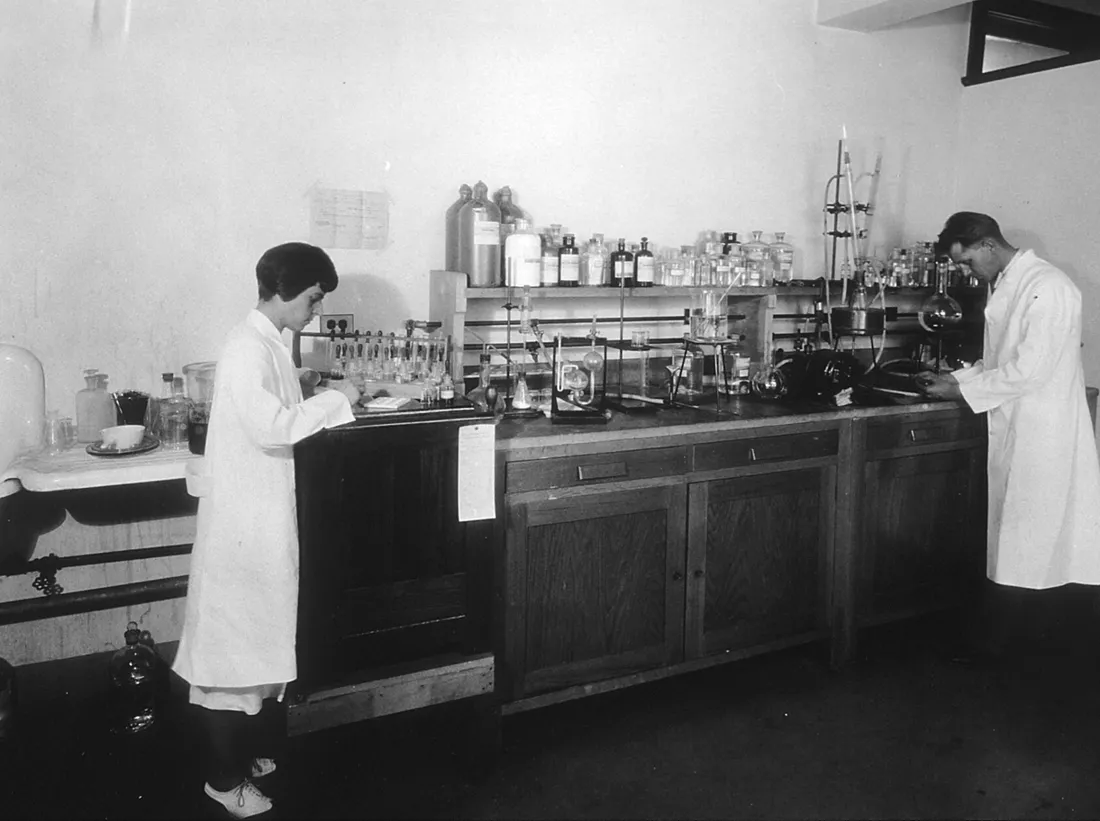 | Sugar chemistry room- central cancer research laboratory | Unknown photographer | Public domain ↗ |
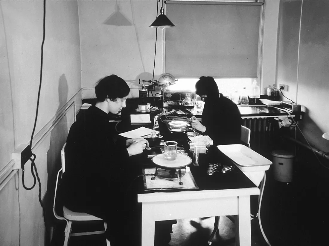 | Tissue culture department at the central cancer research laboratories | Unknown photographer | Public domain ↗ |
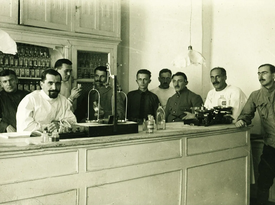 | Scale, pharmacy, pharmacist, interior of a pharmacy Fortepan 85301 | FOTO:Fortepan — ID 85301: Adományozó/Donor: Lőw Miklós. archive copy at the Wayb | CC BY-SA 3.0 ↗ |
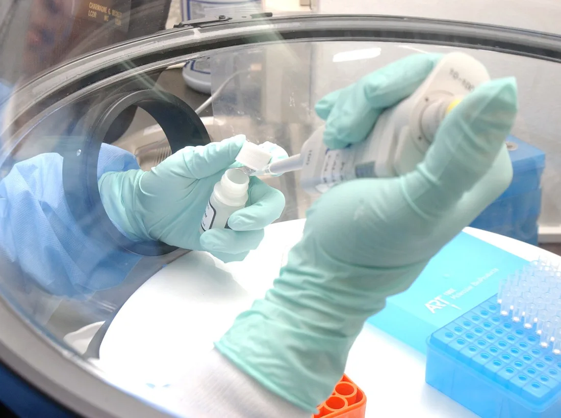 | US Navy 040427-N-0000W-028 Using protective gloves under a protective hood, civilian technician Erlin Listiyaningsih of the NAMRU-2 Laboratory, Jakarta, Indonesia, conducts specialized testing to identify a deadly pneumonia str | U.S. Navy photo by Tom Watanabe. | Public domain ↗ |
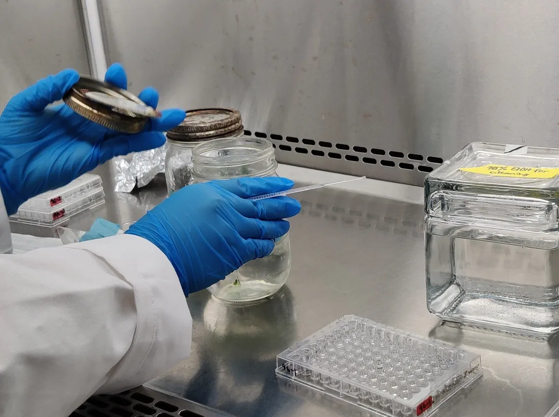 | Scientist At Work | Ns takes photos | CC BY 4.0 ↗ |
 | CSIRO ScienceImage 418 XRay Crystallography Protein Crystals | division, CSIRO | CC BY 3.0 ↗ |
 | Peptide synthesizer | Michael Pereckas from Milwaukee, WI, USA | CC BY-SA 2.0 ↗ |
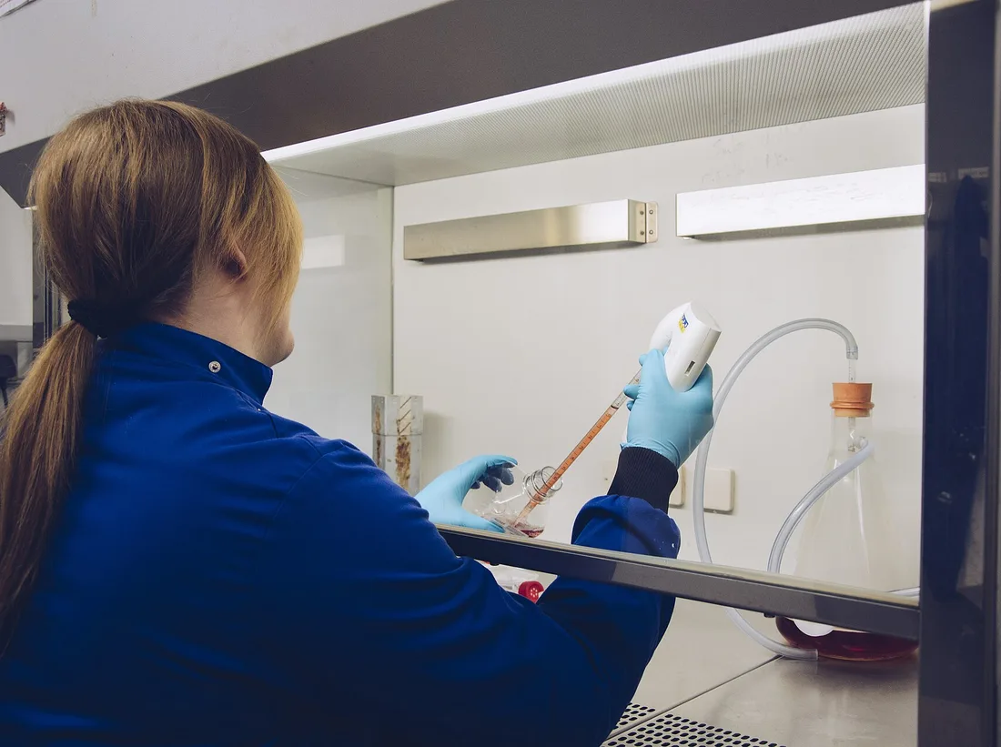 | Tissue Culture | Orla Keogh | CC BY 4.0 ↗ |
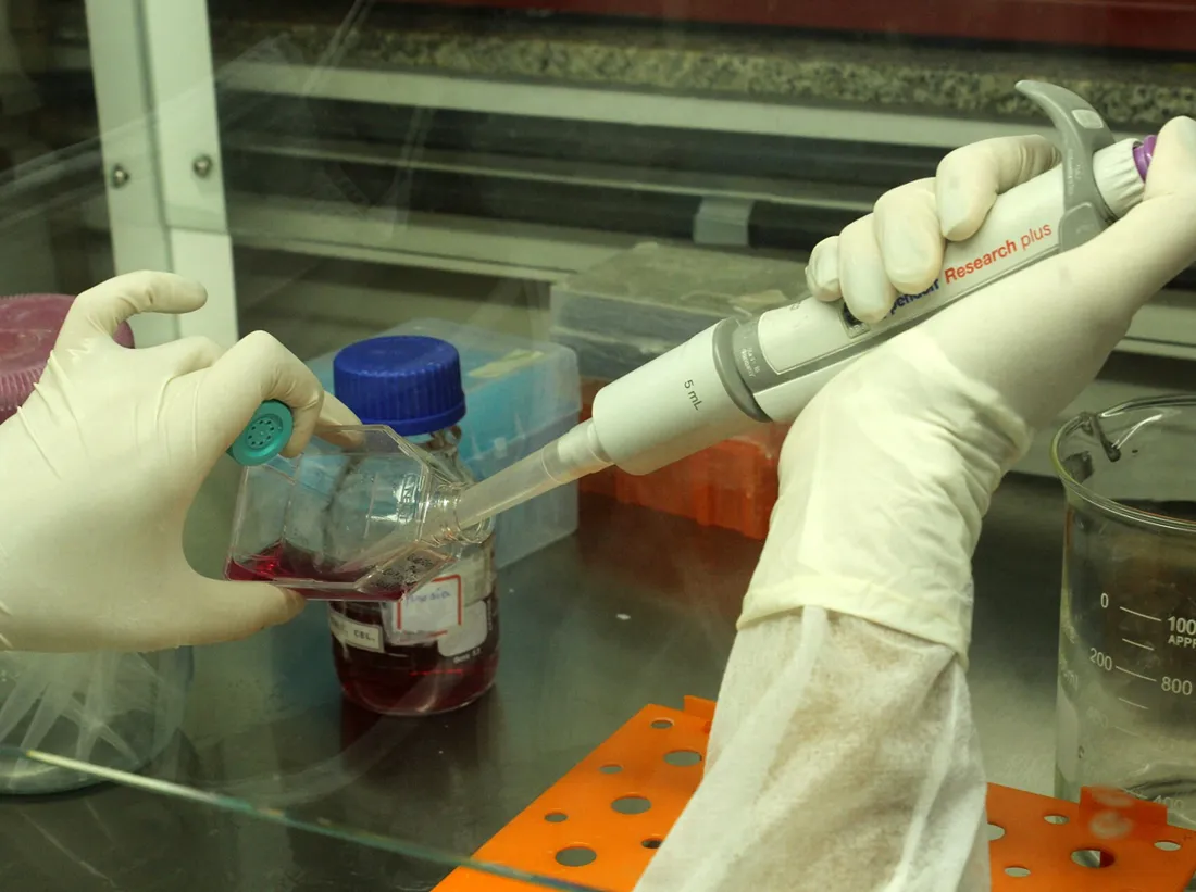 | Biologist studying cancer 04 | Mateus Figueiredo | CC BY-SA 4.0 ↗ |
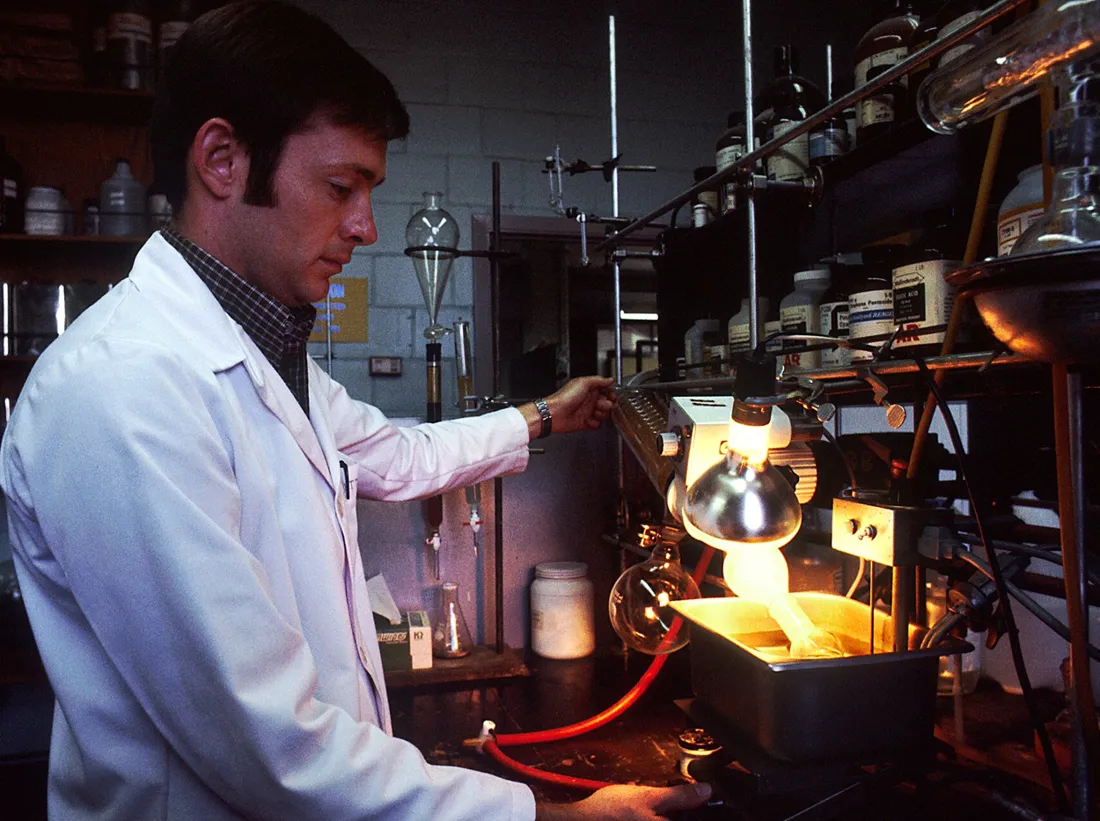 | Laboratory (1) | Linda Bartlett (Photographer) | Public domain ↗ |
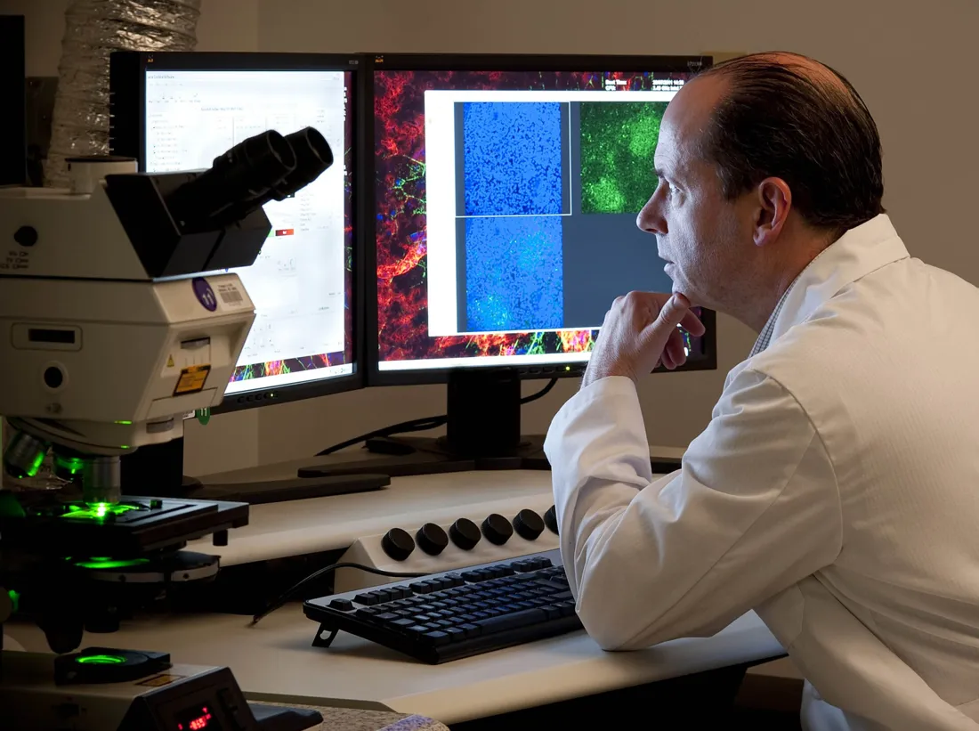 | Observing Ocular Tissue under Laser Scanning Microscope (9955408263) | National Eye Institute | CC BY 2.0 ↗ |
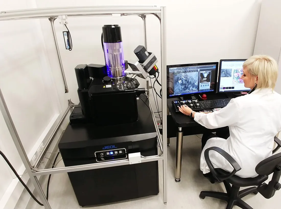 | Scientist during work with Scanning Electron Microscope | Gabriela P. | CC BY 4.0 ↗ |
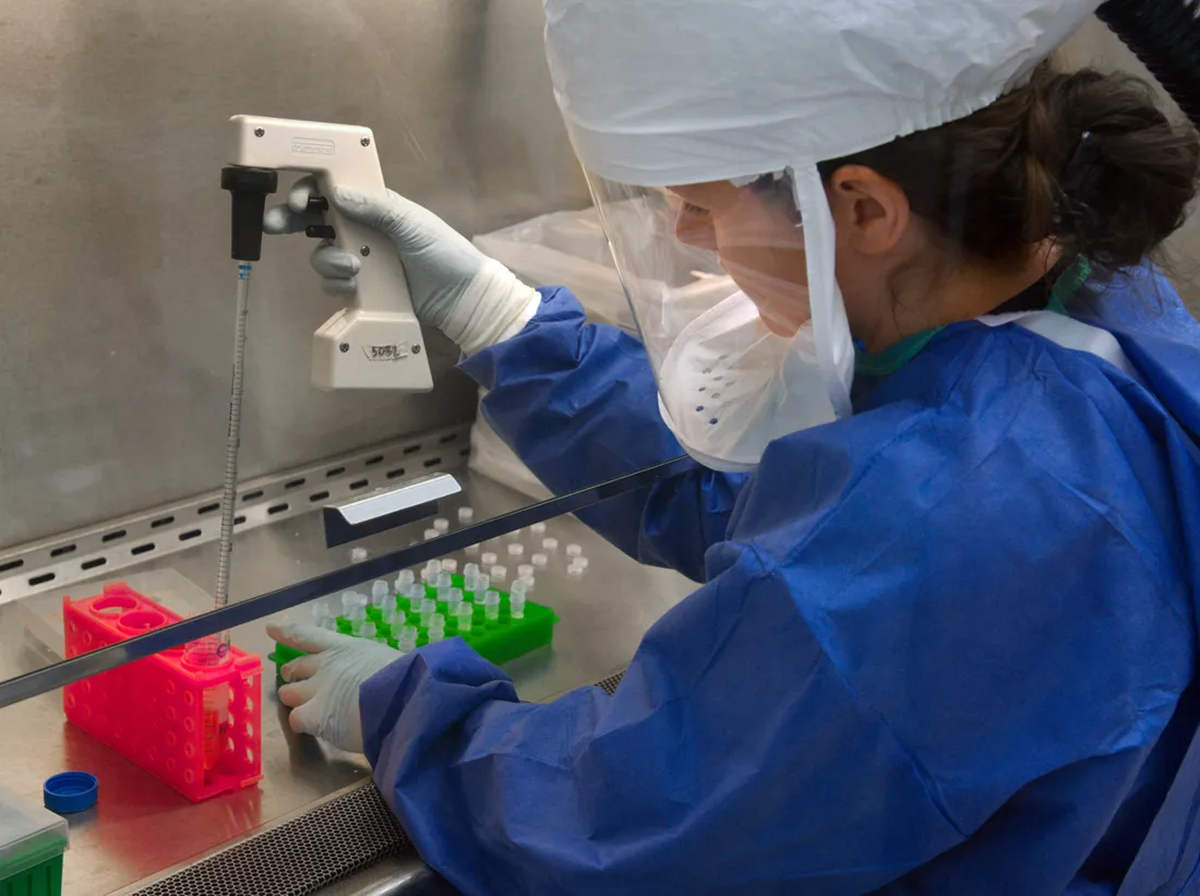 | CDC scientist transfers H7N9 | Photo Credit: James Gathany Content Providers: CDC/ Douglas E. Jordan | Public domain ↗ |
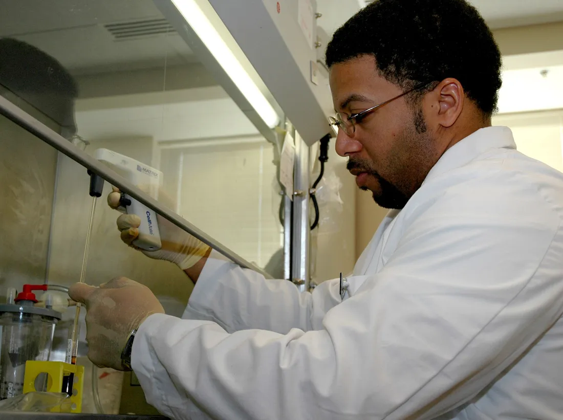 | Pipetting activities | Diane A. Reid (Photographer) | Public domain ↗ |
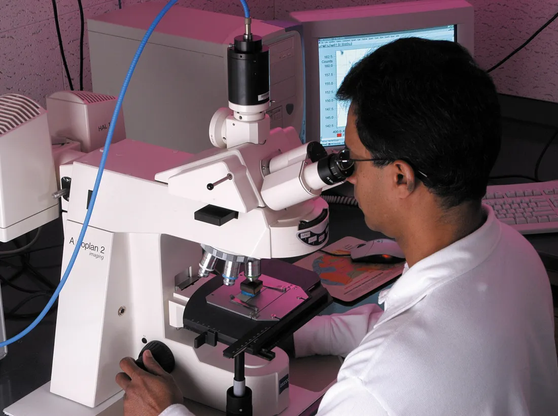 | CSIRO ScienceImage 7845 CSIRO scientist Dr Mohinudeen Faiz analyses a coal sample with methaneproducing microorganisms under the microscope | Chris Taylor, CSIRO | CC BY 3.0 ↗ |
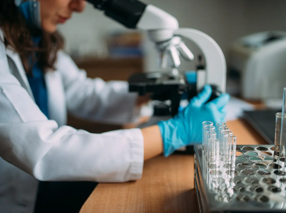 | Doctor looking in the microscope | Nenad Stojkovic | CC BY 2.0 ↗ |
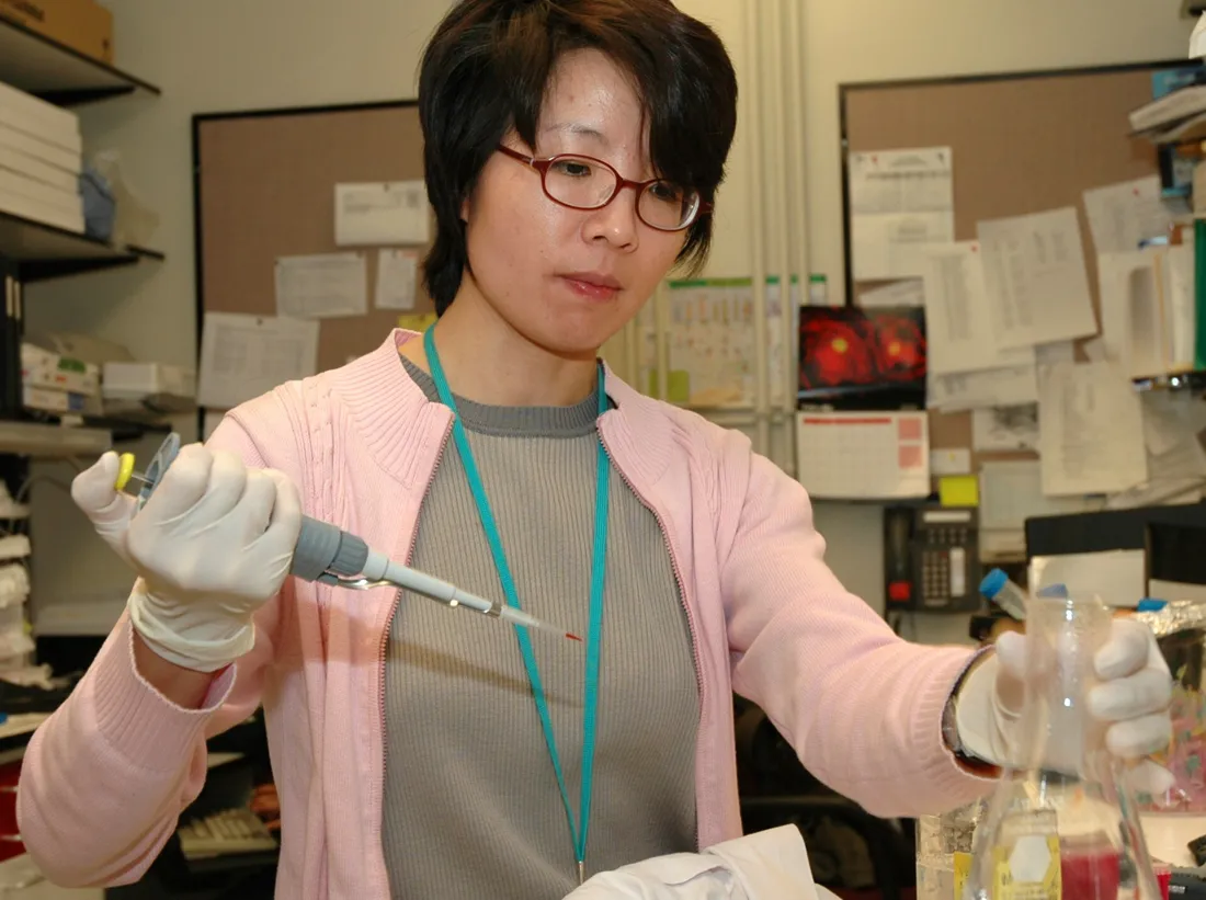 | Pipetting | Diane A. Reid (Photographer) | Public domain ↗ |
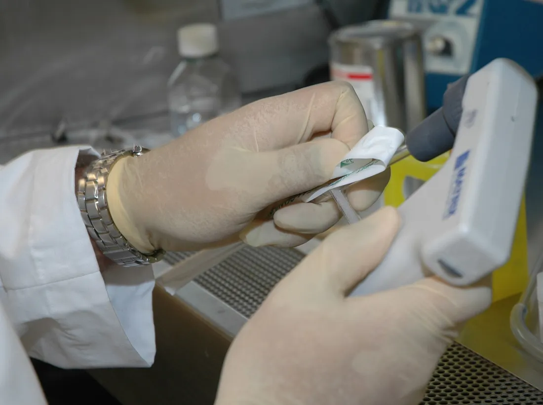 | Pipetting (1) | Diane A. Reid (Photographer) | Public domain ↗ |
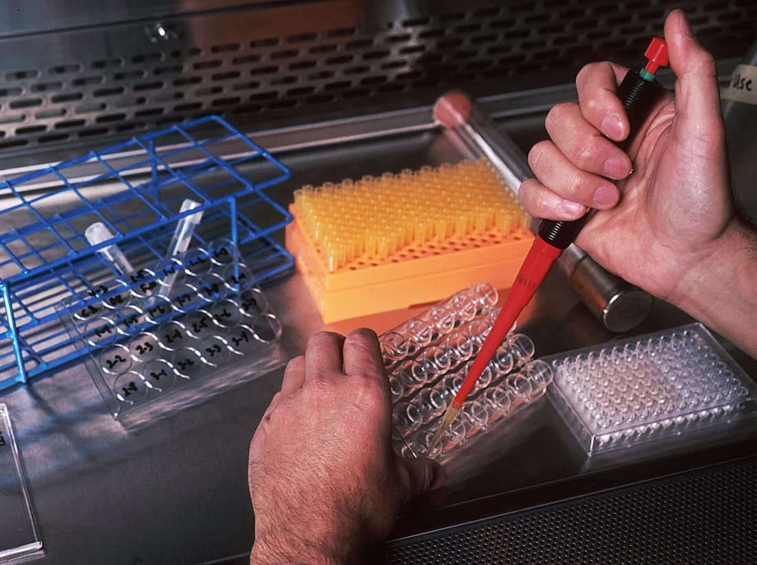 | Monoclonal antibodies (8) | Linda Bartlett (Photographer) | Public domain ↗ |
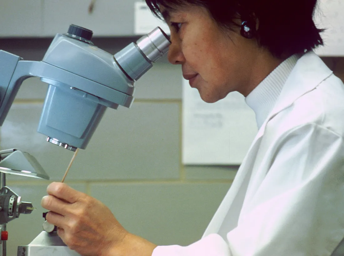 | Scientist looking thorugh microscope | Unknown photographer | Public domain ↗ |
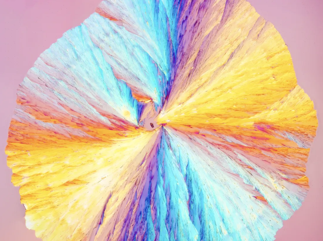 | Recrystallized Amino acid art | Aw1792300 | CC BY 4.0 ↗ |
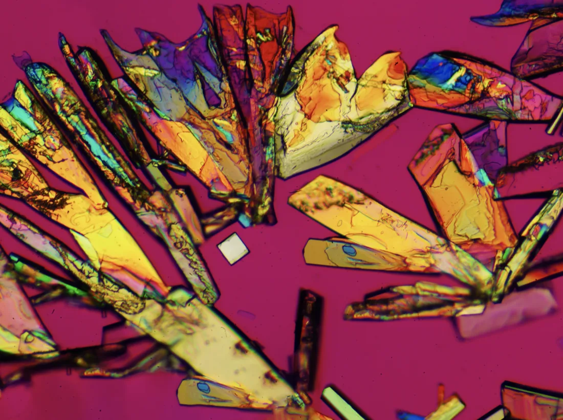 | Recrystallized Amino acid birefringence artwork | Aw1792300 | CC BY 4.0 ↗ |
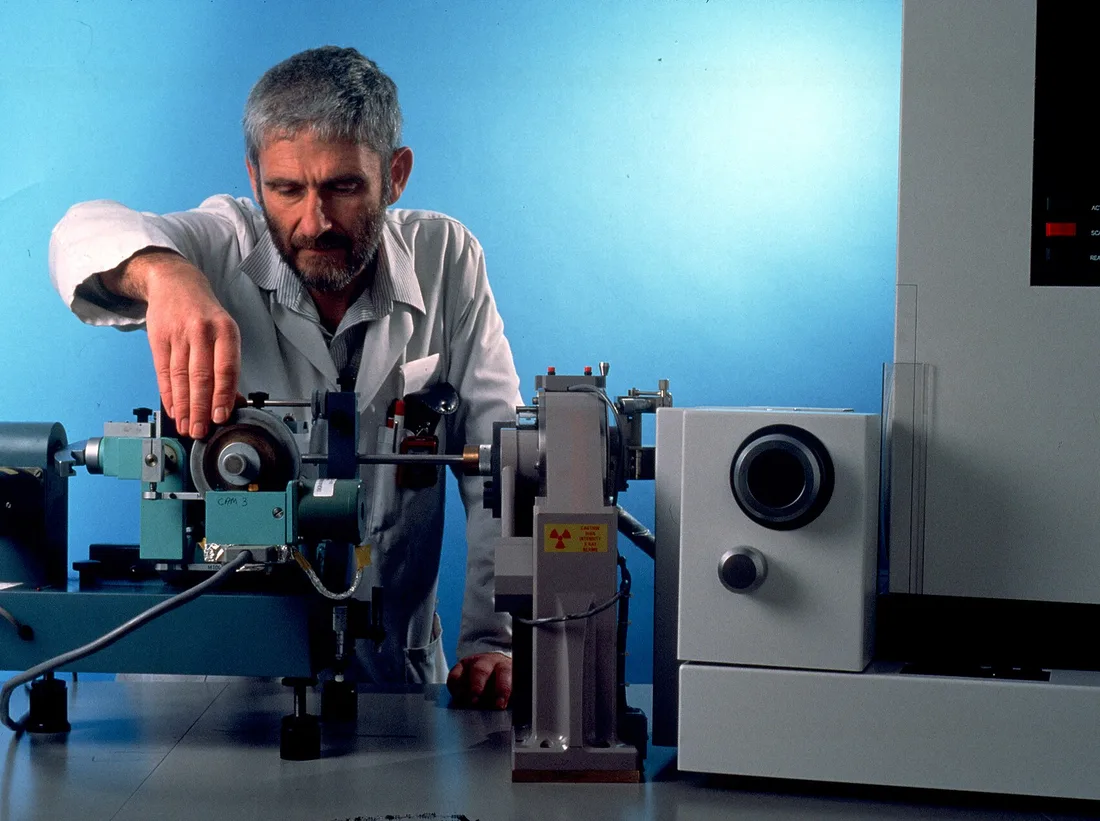 | CSIRO ScienceImage 426 XRay Crystallography Equipment | Health Sciences and Nutrition, CSIRO | CC BY 3.0 ↗ |
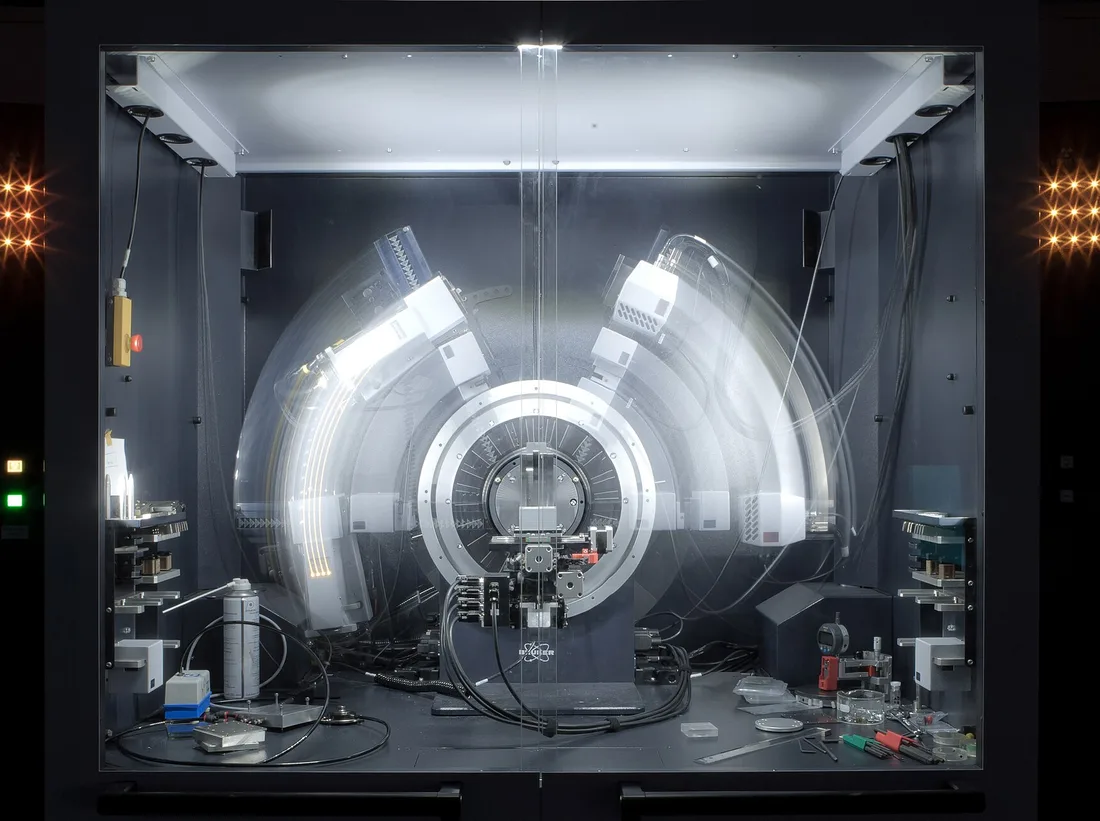 | Freezed XRD | Kaspar Kallip | CC BY-SA 4.0 ↗ |