
Crediti
Crediti fotografici
Fotografie reali provenienti da Wikimedia Commons, archivi del governo degli Stati Uniti e altre fonti di pubblico dominio o con licenza aperta.
| Foto | Autore | Licenza | |
|---|---|---|---|
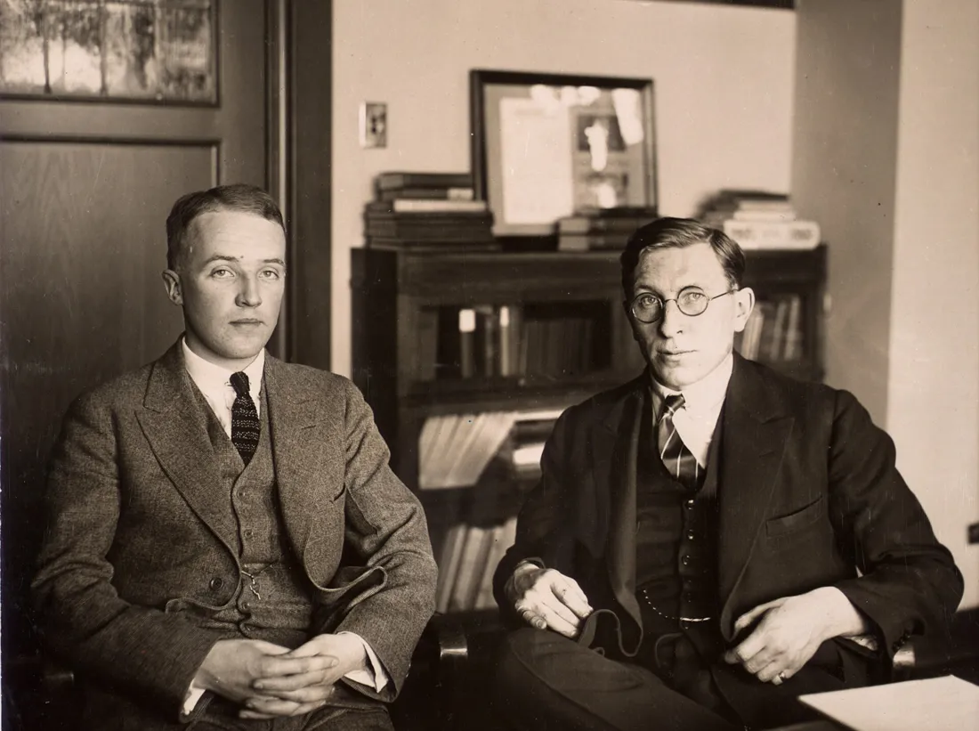 | C. H. Best and F. G. Banting ca. 1924 Insulin P10103 0001 | Unknown author | Public domain ↗ |
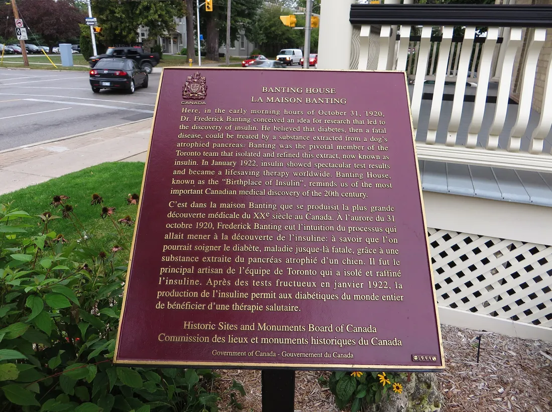 | Banting House, Birthplace of Insulin, London, Ontario (21650793378) | Ken Lund from Reno, Nevada, USA | CC BY-SA 2.0 ↗ |
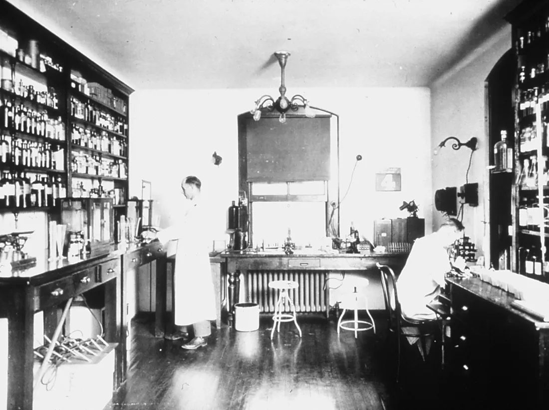 | Kinyoun's laboratory | Unknown photographer | Public domain ↗ |
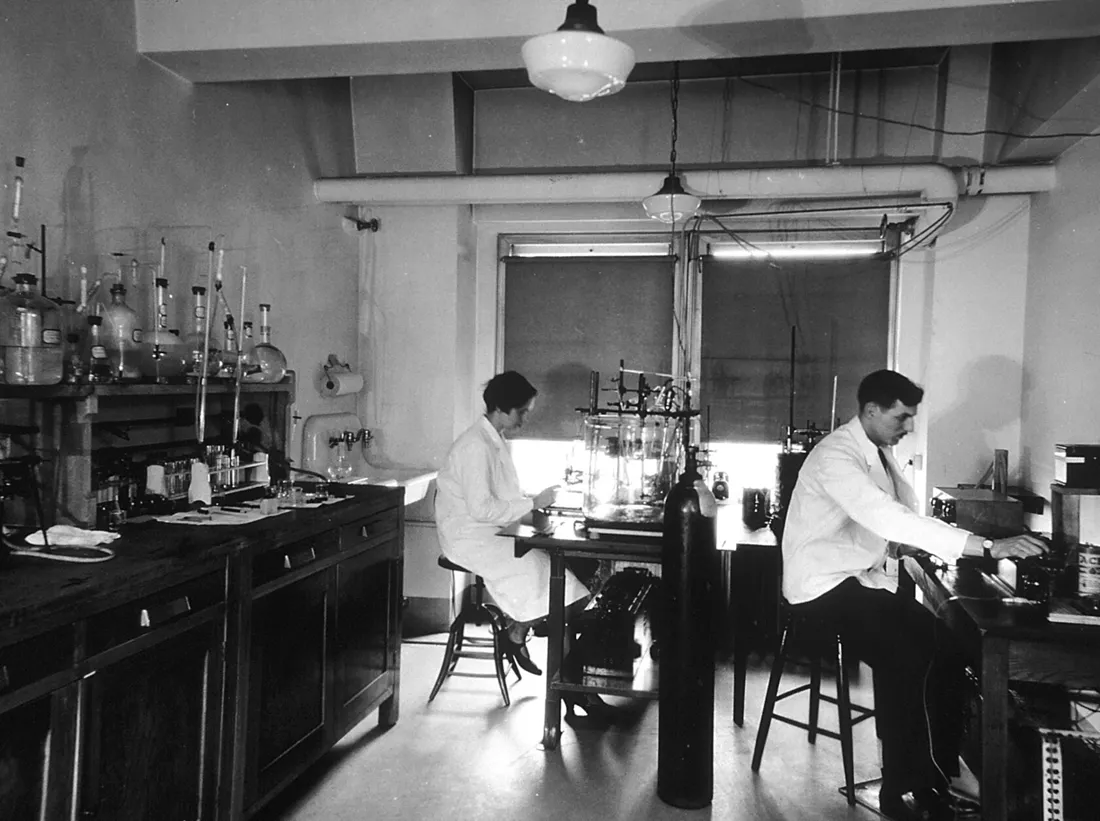 | Chemical laboratory at the central cancer research labs | Unknown photographer | Public domain ↗ |
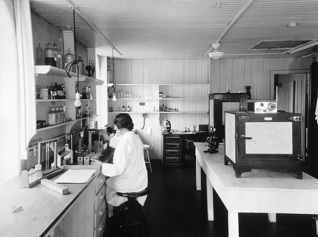 | Immunology laboratory at the central cancer research labs | Unknown photographer | Public domain ↗ |
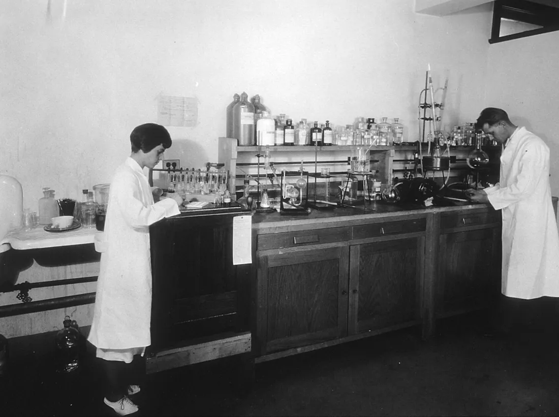 | Sugar chemistry room- central cancer research laboratory | Unknown photographer | Public domain ↗ |
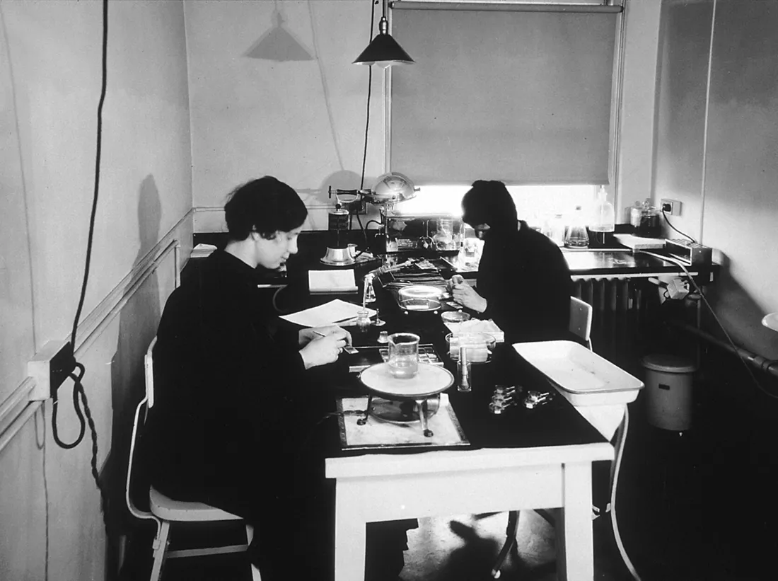 | Tissue culture department at the central cancer research laboratories | Unknown photographer | Public domain ↗ |
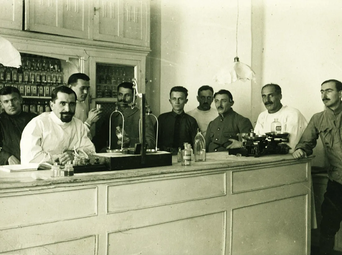 | Scale, pharmacy, pharmacist, interior of a pharmacy Fortepan 85301 | FOTO:Fortepan — ID 85301: Adományozó/Donor: Lőw Miklós. archive copy at the Wayb | CC BY-SA 3.0 ↗ |
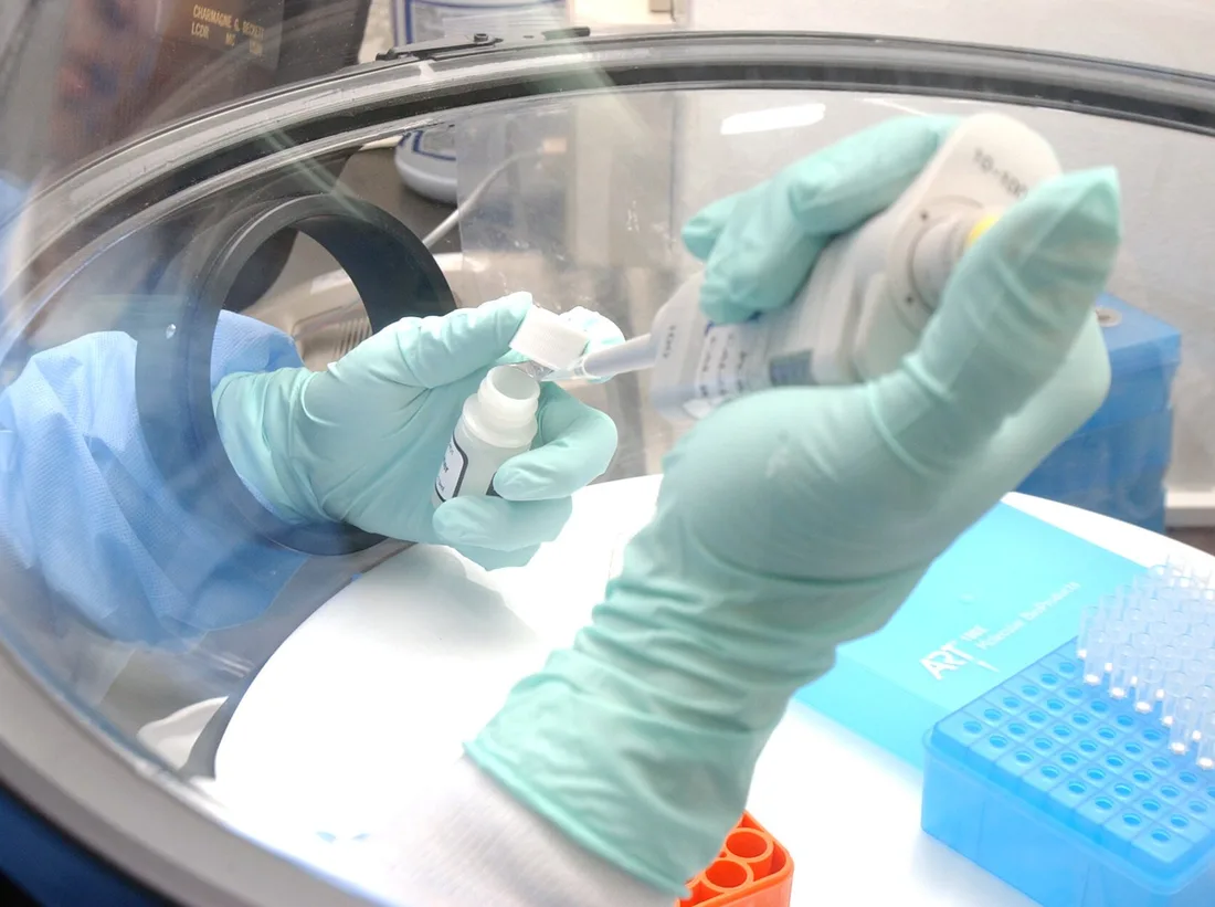 | US Navy 040427-N-0000W-028 Using protective gloves under a protective hood, civilian technician Erlin Listiyaningsih of the NAMRU-2 Laboratory, Jakarta, Indonesia, conducts specialized testing to identify a deadly pneumonia str | U.S. Navy photo by Tom Watanabe. | Public domain ↗ |
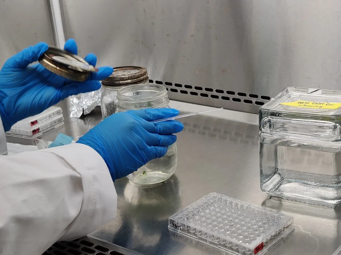 | Scientist At Work | Ns takes photos | CC BY 4.0 ↗ |
 | CSIRO ScienceImage 418 XRay Crystallography Protein Crystals | division, CSIRO | CC BY 3.0 ↗ |
 | Peptide synthesizer | Michael Pereckas from Milwaukee, WI, USA | CC BY-SA 2.0 ↗ |
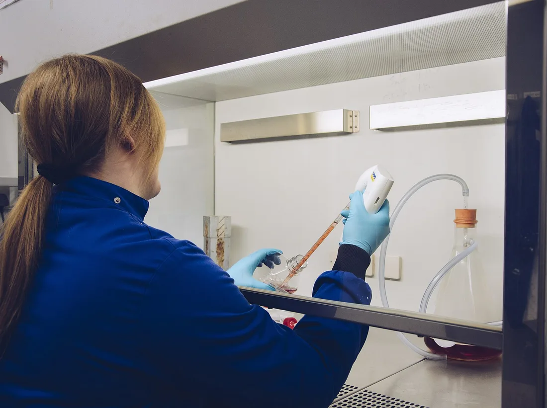 | Tissue Culture | Orla Keogh | CC BY 4.0 ↗ |
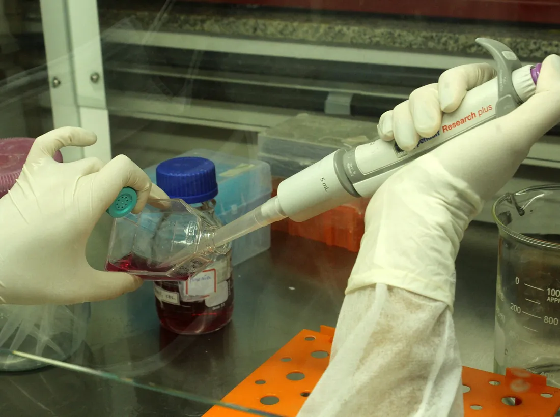 | Biologist studying cancer 04 | Mateus Figueiredo | CC BY-SA 4.0 ↗ |
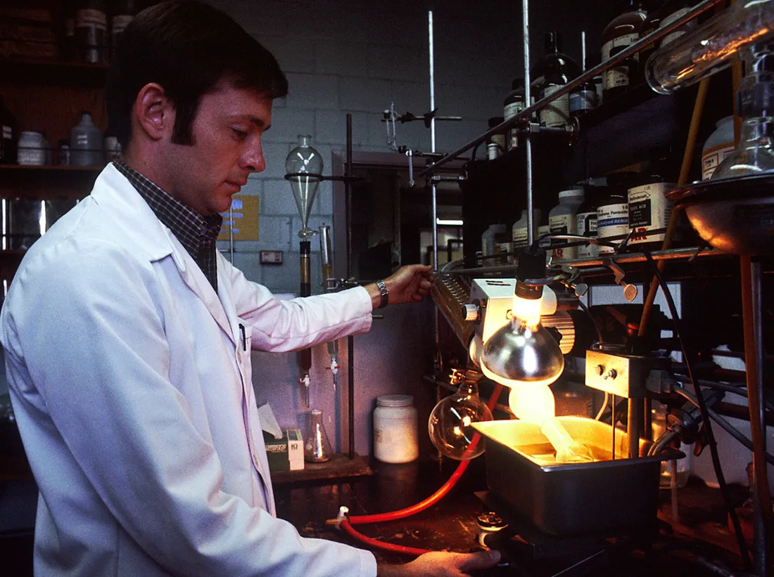 | Laboratory (1) | Linda Bartlett (Photographer) | Public domain ↗ |
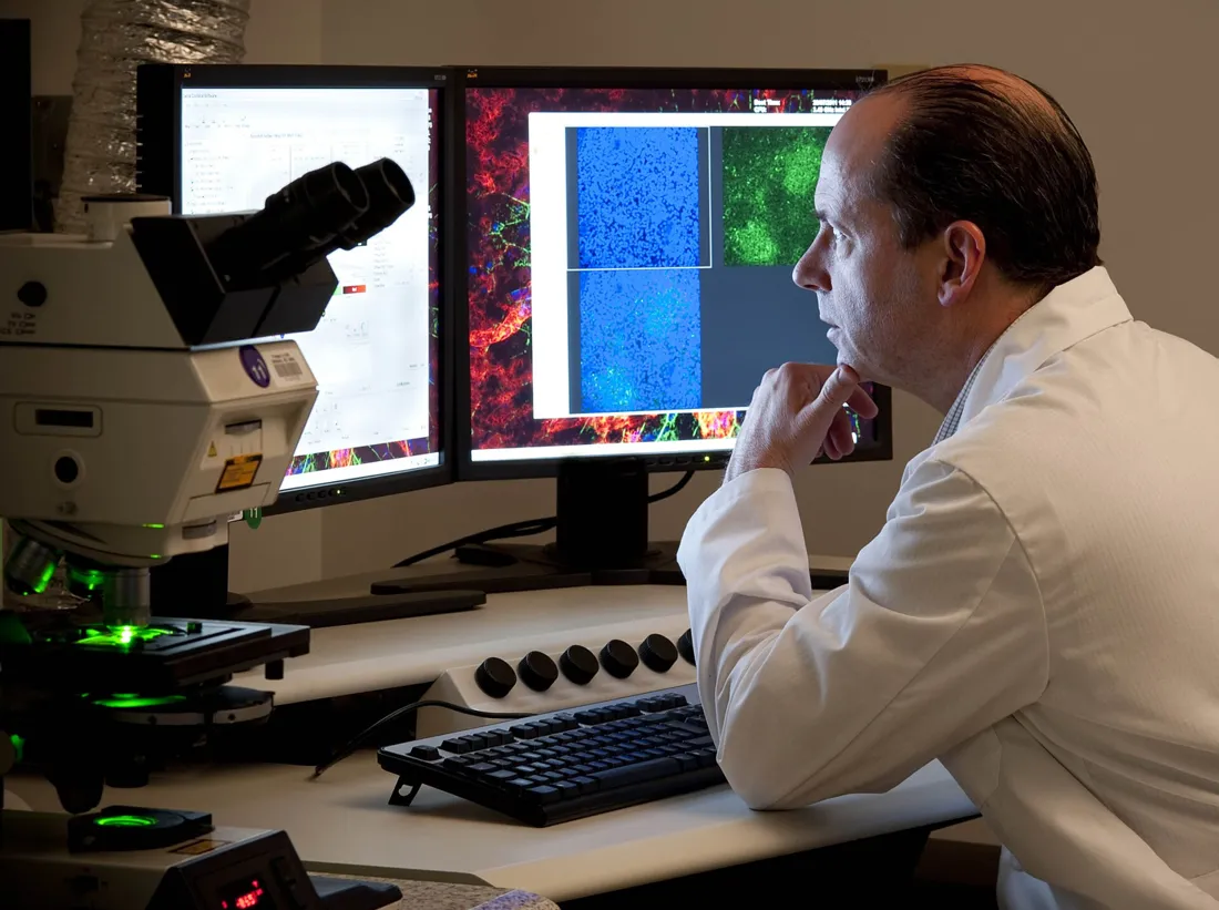 | Observing Ocular Tissue under Laser Scanning Microscope (9955408263) | National Eye Institute | CC BY 2.0 ↗ |
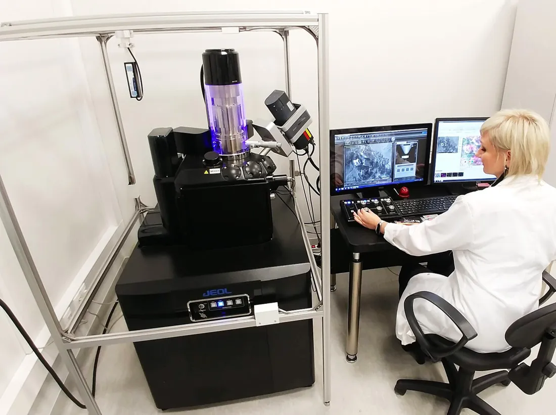 | Scientist during work with Scanning Electron Microscope | Gabriela P. | CC BY 4.0 ↗ |
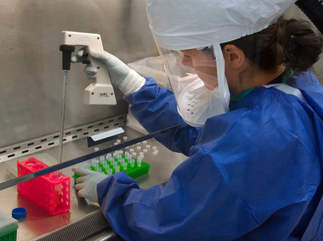 | CDC scientist transfers H7N9 | Photo Credit: James Gathany Content Providers: CDC/ Douglas E. Jordan | Public domain ↗ |
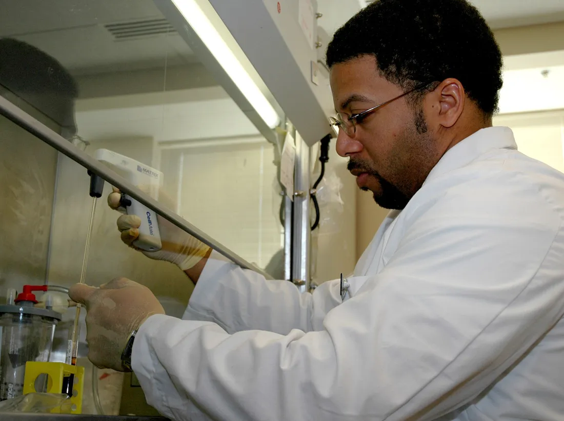 | Pipetting activities | Diane A. Reid (Photographer) | Public domain ↗ |
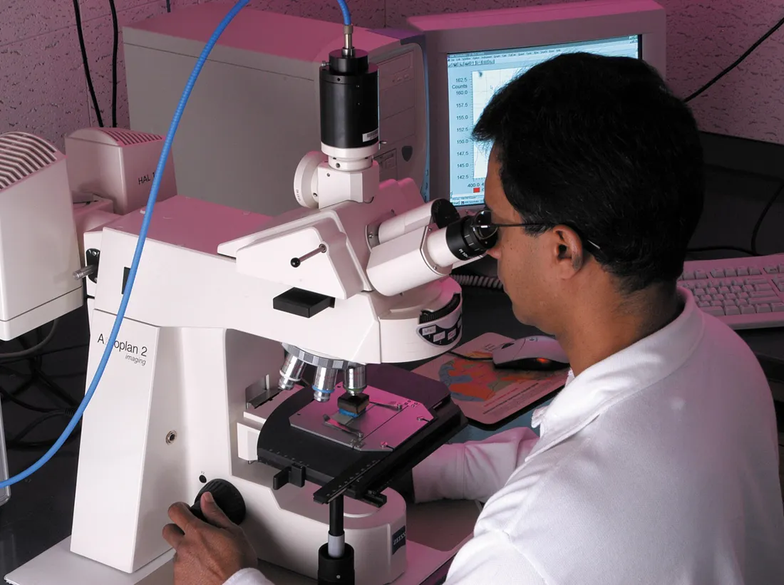 | CSIRO ScienceImage 7845 CSIRO scientist Dr Mohinudeen Faiz analyses a coal sample with methaneproducing microorganisms under the microscope | Chris Taylor, CSIRO | CC BY 3.0 ↗ |
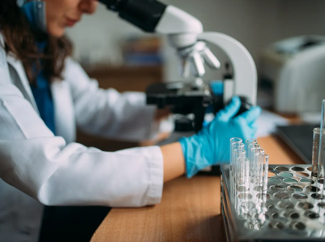 | Doctor looking in the microscope | Nenad Stojkovic | CC BY 2.0 ↗ |
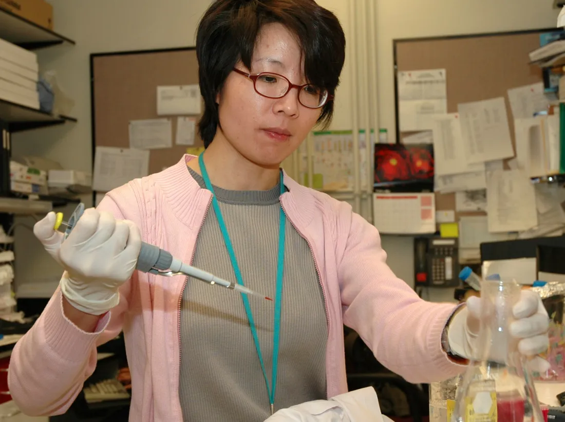 | Pipetting | Diane A. Reid (Photographer) | Public domain ↗ |
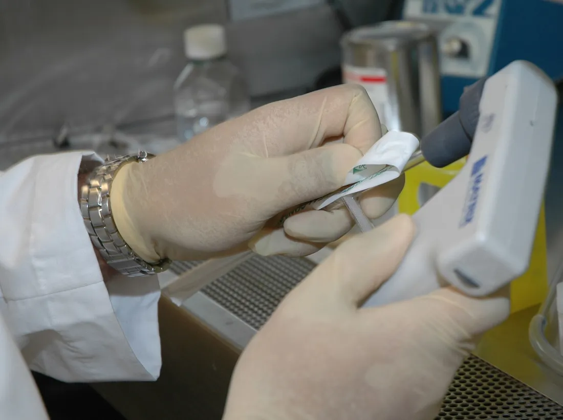 | Pipetting (1) | Diane A. Reid (Photographer) | Public domain ↗ |
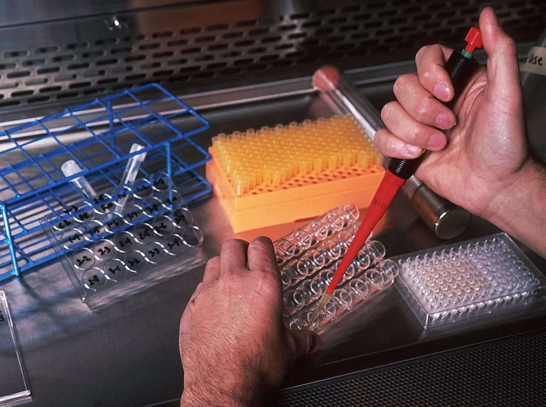 | Monoclonal antibodies (8) | Linda Bartlett (Photographer) | Public domain ↗ |
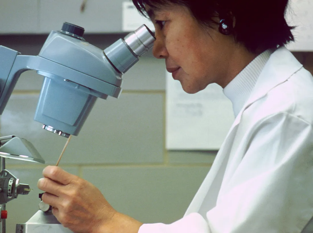 | Scientist looking thorugh microscope | Unknown photographer | Public domain ↗ |
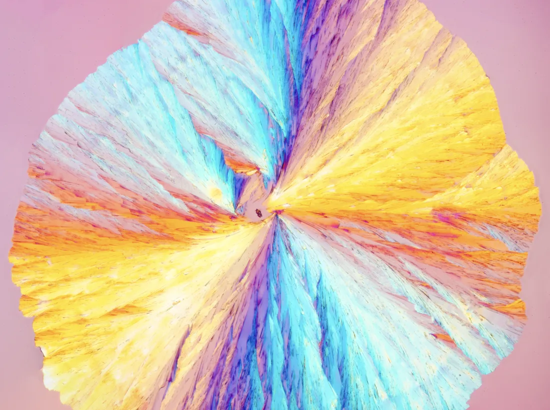 | Recrystallized Amino acid art | Aw1792300 | CC BY 4.0 ↗ |
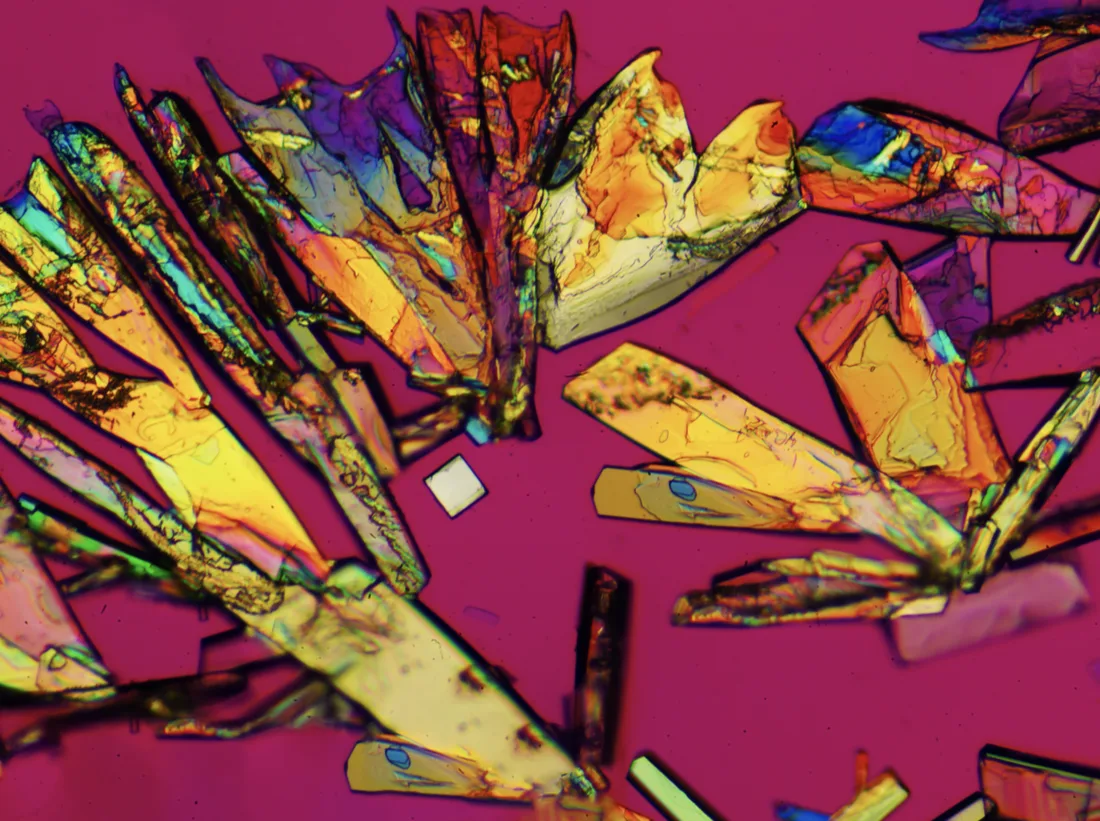 | Recrystallized Amino acid birefringence artwork | Aw1792300 | CC BY 4.0 ↗ |
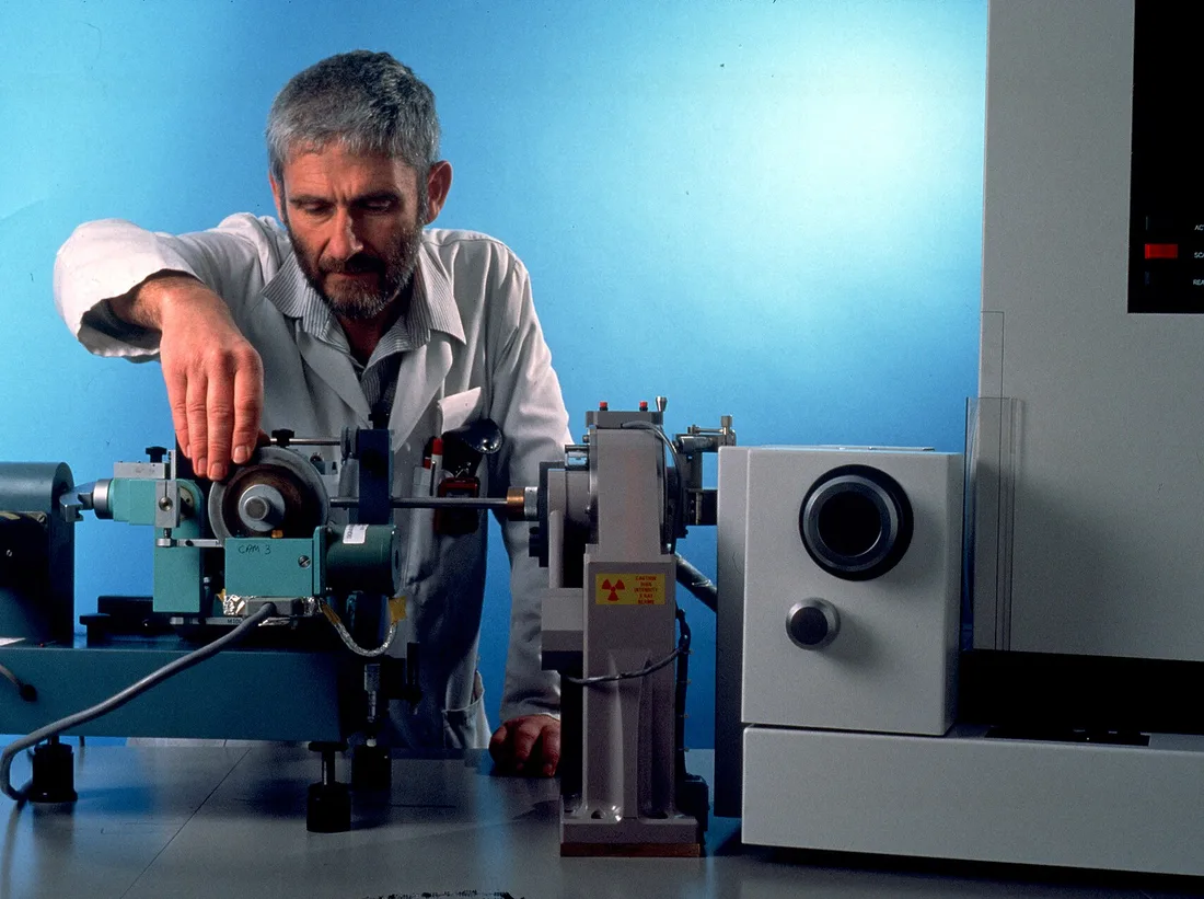 | CSIRO ScienceImage 426 XRay Crystallography Equipment | Health Sciences and Nutrition, CSIRO | CC BY 3.0 ↗ |
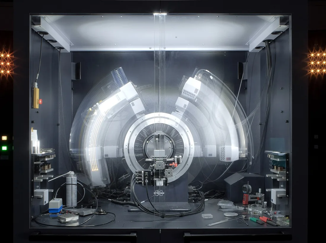 | Freezed XRD | Kaspar Kallip | CC BY-SA 4.0 ↗ |